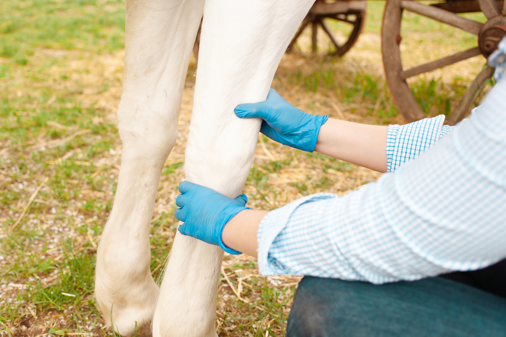
Diagnostic Lameness Exam

Every lameness exam is not the same, thus there are many components to a complete lameness exam.
History
The history of the lameness is very important. How long has the problem been present? What has been tried? What has worked and not worked? Past lameness history and present medication the horse may be receiving.
Visual Exam
We will look at the overall picture of the horse, its conformation, shoeing, muscle symmetry and tone. Palpation of the horse’s legs, joints and musculature is extremely important.
Observing Movement
We may watch them in hard or soft ground. Just on a lunge line or with a rider. How they transition between the gaits maybe very helpful in determining what is going on.
Flexion Tests
This allows us to apply stress to a joint or joints to help isolate were the pain is coming from on the horse.
Joint Or Nerve Blocks
An anesthetic is either injected around a specific nerve or in a joint to help isolate the lameness. It is very important to isolate the localized region that the pain is coming from. Once the area is isolated, we can begin to determine which structure, or structures, is causing pain and creating an abnormal gait.
Diagnostic Imaging
Once the lameness is localized imaging will need to be performed to hopefully identify the cause of the lameness. Imaging may be radiographs, ultrasound, fluoroscope, MRI or nuclear scintigraphy. Depending on the area and type of injury multiple imaging modalities may need to be used to identify the cause of the lameness.
More On Diagnostic Imaging

